In a groundbreaking study, scientists have successfully captured the mesmerizing moment when a zebrafish heart starts beating. The researchers, led by Harvard University biophysicist Adam Cohen, used high-speed microscope imaging to observe the development of zebrafish embryos in detail. This achievement is particularly remarkable considering the short lifespan of a zebrafish. Through their observations, the researchers were able to uncover the intricate process of heart formation in these tiny creatures.
Prior to this study, scientists had already conducted extensive research on the first heartbeats of various animals, including chickens, rats, and mice. It was known that the first heartbeat occurs before the formation of the heart structure itself. The initial signs of activity are visible within populations of heart muscle cells, called cardiomyocytes, which are surrounded by calcium ions. However, the calcium ions are not organized in any specific manner at this stage.
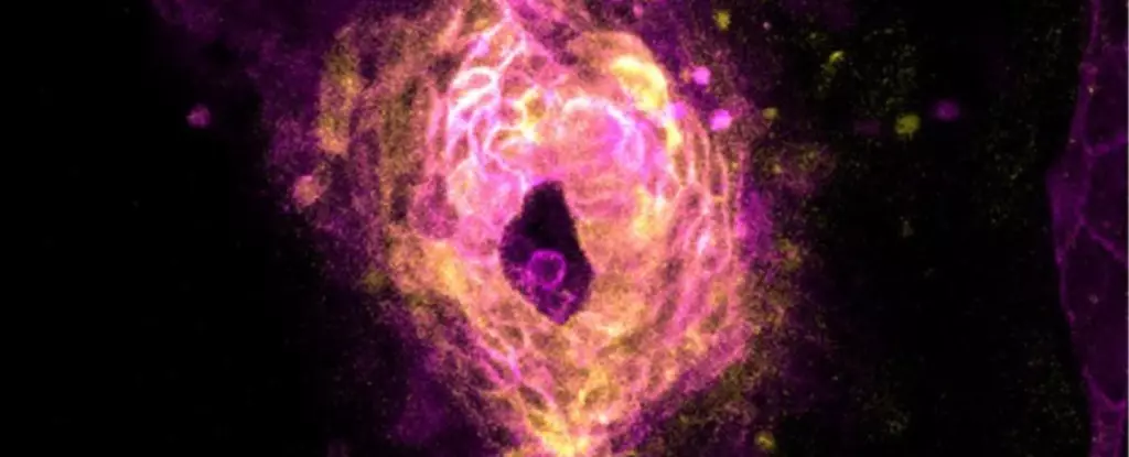
Building upon previous studies, Cohen and his team focused on the development of zebrafish embryos. They closely monitored the waves of calcium ions within the cardiomyocytes and observed how they gradually became more organized and frequent. The cells arranged themselves into a ring shape at the center of the embryos. Suddenly, there was a spike in calcium levels, and the heart cells released bursts of electrical activity, triggering the first few irregular heartbeats. Over time, the contractions became synchronized and the rhythm became more structured.
Interestingly, the researchers noticed that the zebrafish heart cells entered an excitable state approximately 90 minutes before the first heartbeat. This finding suggests that the early activity in heart cells may play a significant role in cardiovascular development. The study also found that the waves of calcium ions preceding the first heartbeat did not consistently originate from the same location within different zebrafish embryos. This indicates that no particular cells have a unique role in initiating the heartbeat. The locus of initiation most commonly occurred in the central region of the cardiac ring, rather than at its outer edges where pacemaker cells reside.
The similarities between zebrafish and other vertebrates, such as chicks, rats, and mice, suggest that the mechanisms of heart formation might be shared across all vertebrate species, including humans. If this is the case, the study could lead to further research into how cardiovascular irregularities, such as arrhythmias, develop in humans. Understanding the intricacies of heart formation in zebrafish could provide valuable insights for improving human health.
The study of zebrafish heart formation has shed light on the mesmerizing process by which the heart begins to beat. The researchers’ innovative use of high-speed microscope imaging allowed them to capture the intricate moments when heart cells organize, triggering the first beats. This groundbreaking research not only deepens our understanding of cardiovascular development in zebrafish but also raises intriguing questions about the similarities across vertebrate species. Further exploration of these shared mechanisms may hold the key to unlocking vital knowledge about human heart formation and potential treatments for cardiac irregularities.
Leave a Reply